(written with help from Ravithree Senanayake)
Leading up to our Fall 2023 CSN All-Hands meeting in Milwaukee, Wisconsin, a group of CSN students, post-docs, and faculty arrived at Deer Creek Intermediate School just outside Milwaukee in St. Francis, Wisconsin. We were greeted by a gaggle of sixth, seventh, and eight graders, all excited for a deviation from their normal school day.
We set up four stations to introduce the students to different aspects of nanotechnology.

Bulk vs. nano
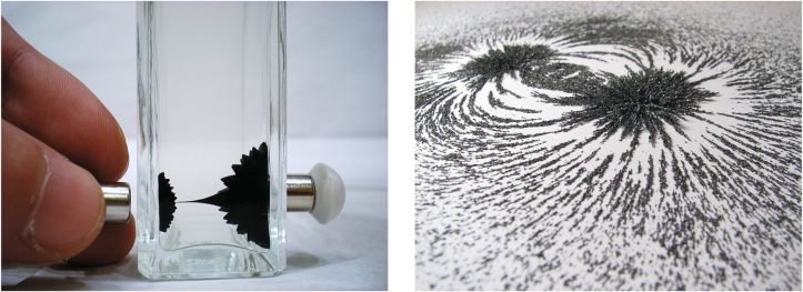
First up, the students were introduced to the difference between bulk materials and their nano-sized counterparts using ferrofluid as an example. Ferrofluid is composed of nano-sized (10 nm or less) iron particles suspended in a carrier (most often water or oil). When exposed to even a weak magnet, ferrofluid forms distinct “spikes” because it wants to find the most stable arrangement with the minimum amount of total energy in the magnetic field. The unique response in the presence of a magnetic field is a direct result of their nano-size. The same material in the bulk state – for our demo we used iron filings – do not form the same distinct spiky shape in the presence of a magnet. In fact, if you put iron filings in the same carrier fluid as nano-iron, they attract much more slowly.
The second example we used to show different properties of bulk vs nano particles was gold. Because of the way they reflect light, gold nanoparticles look red or purple rather than gold! (You can read all about why in this blog post.)

Microscopy
At this station, students learned about TEM, or transmission electron microscopy. We used a demo developed by CSN alum Natalie Hudson-Smith using a PVC pipe and a UV light bulb (a “blacklight”). Although it is common for scientists to use TEM to characterize nanoparticles, the equipment costs a lot and needs a lot of training to use, so it’s not readily available to students outside of research settings. This demo is easy, inexpensive, and fun to use!
For this demo, a UV light is shown on objects of interest, and depending on the material, the light is either blocked by the object or passes through and leaves a signature image on the cyanotype paper below. Since TEM (and this demo) only gives 2D images, some nanomaterials are easy to identify and characterize, while others, depending on the shape, can be tricky to figure out. The demo allows students to experience this challenge in real life. Objects such a bottle tabs and paper clips are easy to identify, while things like marbles and coins, which have less unique 2D “shadows”, are more difficult (but not impossible) to work with.

Check out the paper1 or this earlier blog post to learn more about the specifics of this demo and how you can set it up in your own classroom.
What is nano?
Our third station included a few different components. First, we explained “nanoscale” by showing the size of a nanoparticle relative to the thickness of a strand of human hair.

There was also a “trivia board” that had questions on little wooden doors that opened to show the answers. Questions covered topics such as how nanoparticles were used many years ago for the color in stained glass, and how it is used in our everyday lives today (for example, silver nanoparticles used in socks to prevent odor and new nanomaterials used in bicycles to reduce their weight). They also showed how nanomaterials already exist in nature – for example, how gecko lizards use nano to attach and detach their feet while climbing walls.
Finally, for a fun activity, we distributed some Styrofoam balls, pipe cleaners, and toothpicks to represent NP cores vs. the coatings and coronas that often stick to their surfaces (sometimes by design and sometimes accidentally after interacting with a cell or other biological interface). The students enjoyed making their own innovative NPs with different ligands and coronas! This approach was helpful to explain the structure of a nanoparticle and relate to the corona research studies we do in CSN.
Fluorescent LEGOs
The final station used LEGOs to demonstrate how some researchers make nanoparticles fluorescent in order to help find them in microscopic images. The students were given a big bin full of LEGOs of all different colors and were tasked with counting how many of each color were present. With so many different colors, it was difficult to count each one. The LEGOs were then placed under a UV light, and one color brightly fluoresced (glowed), making those bricks stand out from the rest and very easy to count. This same concept is applicable to certain nanoparticles, as their fluorescent properties can be used to track their movements through things such as microorganisms or plants.


The CSN students, post docs, and faculty and the Deer Creek intermediate School students had a ton of fun at this event. Keep an eye out for more CSN outreach events coming soon!
REFERENCES
- Hudson-Smith et al. A Macroscale Model for Hands-On Activities Demonstrating Transmission Electron Microscopy. Journal of Chemical Education, 2019, 96(7) 1377-1382. DOI: 10.1021/acs.inorgchem.8b01855

