Welcome to the Sustainable Nano library of TEM images (Transmission Electron Microscopy) images. Here, you will find various TEM micrographs to be used for teaching purposes. For our posts on TEM, check the tag here.
 Figure 1, 2. Sample of the bacteria Acinetobacter baylyi exposed to gold nanoparticles. (Credit: Joseph Buchman)
Figure 3, 4. Samples of the bacteria Azotobacter vinelandii. (Credit: Joseph Buchman)
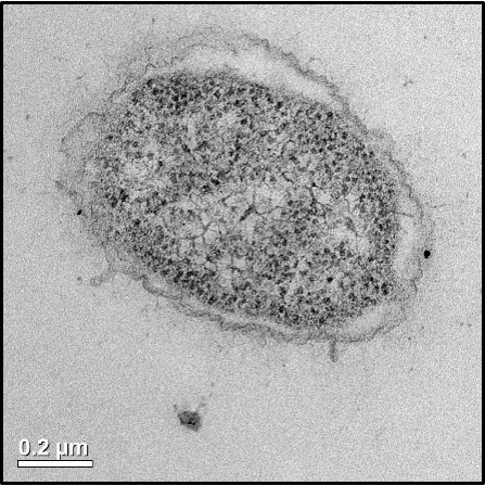
Figure 5. Samples of the bacteria Shewanella oneidensis MR-4 exposed to gold nanoparticles. (Credit: Joseph Buchman)
 Figure 6, 7. (left) Shewanella oneidensis MR-1 exposed to phosphorous doped silicon nanoparticles. (right) the same frame in dark-field mode. (Credit: Bo Zhi)
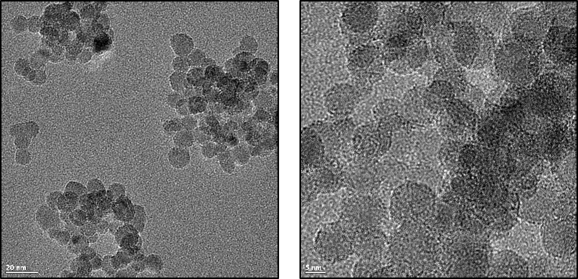 Figure 8, 9. Silicon nanocrystals doped with 30% boron. (Credit: Bo Zhi)
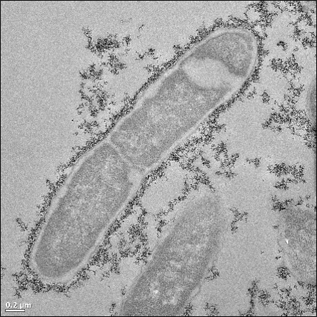 Figure 10. Shewanella oneidensis MR-1 exposed to silicon nanocrystals doped with 30% boron. (Credit: Bo Zhi)
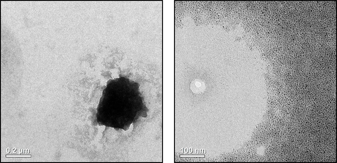 Figure 11, 12. Unidentified debris observed on TEM preparation grids. (Credit: Natalie Hudson-Smith)
 Figure 13, 14, 15. Salt crystals co-localized with a sample of copper nanoparticles. (Credit: Natalie Hudson-Smith)
 Figure 16, 17. Carbon dots made from citric acid and urea in molar ratio of 1:1 and synthesized by microwave assisted hydrothermal treatment. (Credit: Xiaoxiao Yao)Parts of this work were carried out in the Characterization Facility, University of Minnesota, which receives partial support from NSF through the MRSEC program .

