In the 1930s, microscope designer Royal Rife made a splash with reports that he had designed a new microscope that could view nanoscale objects such as viruses!1 The only problem was that it didn’t work. In fact, it couldn’t work, based on the basic physics of light. Rife was attempting to improve upon the optical microscope (which uses visible light shining through lenses) but the feats he claimed would not be possible except with an electron microscope.
But why?


You might have already noticed how many types of microscopes there are, ranging from simple optical microscopes (like the microscope emoji 🔬) to expensive and complex electron microscopes. There is a litany of electron microscopes with names so long that we just make acronyms – SEM, TEM, STEM* – rendering our lab notebooks into what looks to be an alphabet soup. Electron microscopes are a valuable tool in nanotechnology because they allow us to visualize and understand objects that are on the nanoscale.
So what’s the difference between an electron microscope and a simple optical microscope? Why can’t you just take the microscope from a high school biology lab, slap a couple more lenses on it, crank that magnification up, and check out some nanoparticles? Why can’t we use the same kind of microscope to visualize every kind of sample?
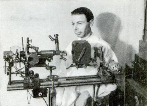
In 1932, a man named Royal Raymond Rife asked the same questions. He claimed to have invented an optical microscope (similar to what you would have in a high school lab) that used visible light to see objects on the nanoscale. Rife seemed like a reputable microscopist – he was employed by Zeiss, a company that still makes microscopes for scientists today.2
However, his inventions never lived up to his claims.
Rife began to design his own microscopes and named them the Rife 1 through 5. Of these microscopes, the Rife 3 was the most famous and most coveted. The Rife 3 was touted as a “Universal Microscope” – one microscope for all your needs. Rife even claimed that this device could resolve images of live and active viruses, which were much too small (~200 nm, or 200 billionths of a meter) to visualize with the existing microscopy techniques at the time.3,4
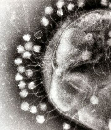
If Rife was telling the truth, this would have been a ground-breaking discovery in microscopy. Modern microscopy techniques can resolve images of viruses like the one above but they require a long and tedious preparation to deactivate and immobilize the virus. These preparations include staining a virus to provide contrast, embedding the stained virus in a plastic resin (like a bug in amber) and slicing thin cross sections of the plastic to examine one by one.5 These processes are so expensive and time-consuming that even modern scientists would be thrilled to have alternative techniques like those Rife promised.
Of course, Rife’s claims generated a flurry of interest among his colleagues and they requested to use the device to observe these amazing discoveries themselves. It was at this point that people started to realize Rife was not representing his microscopes honestly.
Although his microscopes did function as microscopes, they didn’t achieve his claim – the ability to see viruses. To increase magnification, Rife had added many extra lenses to his devices. These lenses slightly increased magnification but greatly reduced resolution and introduced many optical artifacts. (See this post illustrating magnification vs. resolution using dog photos.) It is possible that Rife saw these optical artifacts and genuinely thought they were viruses. Based on my reading, I can’t be certain whether he was deliberately trying to trick people.
Ultimately, the feats of the Rife – that so called “Universal Microscope” – were never replicated by any other scientists. Other researchers who asked for the Rife 3 model microscopes received them with missing parts so they couldn’t even be put together. In the end, Rife used the parts of the only complete Rife 3 to make the newer models of his microscopes, which he never claimed to be Universal. Royal Rife died discredited and penniless. (He did, however, win some speedboat racing competitions and invent a 100 string guitar, so not all was lost!)
Based on our modern understanding of how light diffracts, we know that it would be physically impossible for Rife’s microscopes to work as he claimed. Microscopy is often referred to as a “diffraction limited system” – this means that the resolution of the microscope is limited by the wavelength used to observe an object. (The same is true of a telescope or a camera.) When light waves pass through a small hole (like the aperture of a camera or microscope), they bend as shown in the figure below. This bending of the light waves is called diffraction.
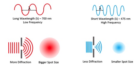
Remember that we see objects when light waves bounce off them and then reach our eyes (for a refresher on this idea, check out this earlier blog post). But the object has to be big enough for the light to bounce off it – the spot sizes represented (not to scale) in the figure above are basically the smallest sizes that each wavelength will bounce off.
So although the magnifying power and resolution of some microscopes are limited by the number and quality of lenses, even the most perfectly crafted microscope would be limited by the wavelength of visible light. We can determine this limit by a simple rule of thumb developed in 1873 – long before Rife made his microscopes – by a physicist named Ernest Abbe.
The Abbe Limit of Diffraction Equation is
d = λ/1.6
where λ (lambda) is the wavelength of our light and 1.6 is a constant representing the shape and quality of the microscope, and d is the spot size made by our diffracted light.6
We can calculate the different spot sizes for different colors in the visible spectrum based on knowing their wavelengths.**

For example, for red light with a wavelength of 700 nm,
d = 700 nm / 1.6
d = 437 nm , which means that anything smaller than 437 nm would not be resolved in a microscope using that red light.
If we do the same calculation for blue light (λ = 475 nm), we would obtain a spot size of 297 nm. Since 297 nm is larger than many viruses (which range from 20-400 nm), this spot size is just too big! A microscope that relies on seeing things with regular visible light simply will not work.
This is like trying to measure the length of a watermelon by looking at your car’s odometer as you drive past! You can try to drive alongside the watermelon from one end to the other, but the circumference of the car tire is much too large to give an accurate measurement of the watermelon length so the odometer would never be able to display the length of a single watermelon. Just as it is impossible (and ridiculous) to measure a watermelon using a vehicle odometer, it is impossible to look at viruses using the wavelength of visible light.
Clearly, visible light isn’t the right tool to study viruses, so what else could we use?
It turns out that electrons (one component of atoms, along with protons and neutrons) have a wavelength (λ) of 1.23 nm. This is much smaller compared to even the shortest wavelengths of visible light!
If we plug that into our Abbe calculation,
d = 1.23 nm / 1.6 = 0.77 nm
we find a spot size of 0.77 nm which is much smaller than our viruses!
From here, we can do even better. Electrons are subject to the limit of their de Broglie wavelength rather than the Abbe limit of diffraction (although we can use Abbe to make a rough estimate). By accelerating electrons, we can achieve wavelengths of 0.01 nm and even smaller spot sizes!
Using electrons instead of visible light, we can resolve things even smaller than a nanometer (one billionth of a meter). High-resolution transmission electron microscopy can achieve resolution of 0.05 nm! This is why many nanoscientists, myself included, make use of electron microscopes to study nanomaterials such as lithium nickel manganese colbalt oxide battery materials (see blog posts here and here) or mesoporous silica nanoparticles (shown below) which have a range of possible medical and technological applications.

There are many different types of electron microscopes and many different techniques within electron microscopy Despite this variety, at the root of all of them is the very simple reason we use electron microscopy at all – the limit of diffraction! And it is this very simple limitation that prevented Rife’s dreams from coming true for the field of microscopy and ultimately kept the Rife 3 out of the history books – one relatively simple law of physics.
A Note to Readers
Although he is an interesting figure in history, Royal Rife is an incredibly difficult person to obtain accurate information on. This is because he also claimed to have invented a cure for cancer (he didn’t) and his “medical work” underwent a pseudoscientific revival in the 1980s. In this blog post, we cover only his microscopy inventions (or lack thereof). Accurate information on his medical research is scarce and difficult to find amongst all the sales pitches for his so-called cure.7
Footnotes
* SEM is Scanning Electron Microscopy, TEM is Transmission Electron Microscopy, AFM is Atomic Force Microscopy, STEM is Scanning Transmission Electron Microscopy. Each of these deserves their own blog post and you can find more about some of them here, here, here, and here.
** The denominator 1.6 that I am using here assumes a most perfect possible microscope – consider these to be the best possible results and not likely to be obtained in any real microscope!
Author’s Note
This post was revised on March 19, 2019 to better reflect the fact that electron waves are not subject to the Abbe limit of diffraction. We thank our readers for pointing out this error. For more on electron microscope resolution, we recommend this page by the University of Oklahoma.
EDUCATIONAL RESOURCES
- Microscopy Society of America: Project Micro middle school classroom activities
- ThermoFisher Scientific: An Introduction to Electron Microscopy
- Discovery Education: Virtual Electron Microscope lesson plan (grades 6-8)
- Scanning Tunneling Microscope video: A Boy and His Atom: The World’s Smallest Movie
REFERENCES
- Wikipedia, Royal Rife (website)
- DaveF. (2002-2005) History of Royal R. Rife, Jr. (and the Rife Ray machine). DFE Research, 2002-2005 (website)
- Seidel, E. & Winter, M. (1944). The New Microscopes. Annual Report of the Board of Regents of the Smithsonian Institution. Smithsonian Institution: 207–216.
- Rosenow, EC (1932). Observations With The Rife Microscope Of Filter-Passing Forms Of Microorganisms. Science. 76 (1965): 192–3. PMID 17795318. doi: 10.1126/science.76.1965.192.
- Laue, M. (2010) Electron Microscopy of Viruses. Methods in Cell Biology: 96, 1-20. doi:10.1016/S0091-679X(10)96001-9
- Wikipedia, Numerical Aperture (website)
- McElroy, J. (2014) Royal Rife’s Cancer-Curing Death Ray. Sawbones (podcast)


[…] Source: Royal Rife’s Universal Microscope (and Why It Can’t Exist) […]
The Abbe limit of diffraction is only valid for far-field optical treatments – Rife’s microscope relied on coupling of evanescent waves to support their propagation and focus in the far field.
Also, in 1944, the Smithsonian Institute examined his microscope and found it to operate as described.
http://rifevideos.com/1944_smithsonian_report_of_the_rife_universal_microscope.html
Thanks for your comment. One goal of this post was to explain why Rife’s claims about his microscope are not physically possible. Unfortunately, in the Smithsonian article you mention, it sounds like they were not able to see the microscope itself or independently verify Rife’s claims — they were only able to cite reports from others. In addition, that report was focused on bacteria rather than viruses, and bacteria are significantly larger than the nanoscale objects we’re discussing in this post.
[…] on August 18, 2017August 18, 2017 by […]