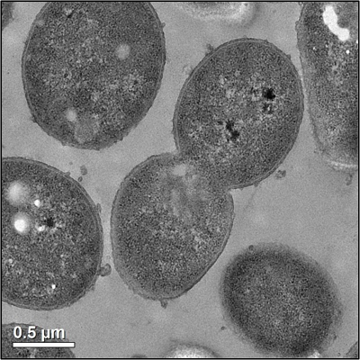
Did you know that under the right conditions, bacteria can divide every 20 minutes?1 (That’s how bacteria reproduce – one cell splits into two.) That means that 10 bacterial cells can produce 2,621,440 bacteria after only 6 hours! No wonder you can get sick so quickly after eating food contaminated with bacteria. Figure 1 below shows one bacterium (a single bacteria) dividing in one of my experiments. But how exactly do bacteria replicate? Let’s dig deeper into that question and then look at one technique scientists in the Center for Sustainable Nanotechnology use in the lab that takes advantage of bacterial replication.
Bacteria reproduce by a process called binary fission. In binary fission (shown in Figure 2), the bacterium first duplicates its DNA, so it has two identical copies of genetic material. The cell then swells and elongates as it gets ready to split, with the copies of DNA moving to separate sides of the cell. A divider then forms in the middle of the elongated cell, which pinches off to form two new cells.

These two new “daughter” bacteria cells are identical because they each have identical copies of DNA from the original bacterium. As I said, bacteria can divide as quickly as every 20 minutes, which means that it doesn’t take long for the bacteria to grow and divide until they’ve formed a cluster of bacteria that is large enough for the human eye to see. These bacterial clusters are referred to as bacterial colonies. You can see a great sped-up video of bacterial cells dividing here.
You may remember that in my last blog post I introduced the Live/Dead® BacLight™ assay, which scientists in the CSN use to determine if bacteria are killed by nanoparticles. Now that you understand how bacteria divide, let’s explore another technique scientists use that takes advantage of the replication rate of bacteria, called drop plate colony counting. This technique was first introduced in 1938 by A. A. Miles and S. S. Misra,2 and is therefore sometimes called the Miles and Misra method. In the CSN, we use colony counting to compare the toxicity of different nanoparticles to a given type of bacterium.
To perform a drop plate colony counting assay, we first expose bacteria to nanoparticles by mixing them together in a liquid suspension. After giving the nanoparticles time to interact with and potentially kill some of the bacteria, a tiny amount of the bacterial suspension is dropped onto an LB agar plate and dried. LB agar is a nutrient-rich medium that allows many organisms, such as bacteria, to easily grow by providing them with all the nutrients they need to thrive. The plates are incubated overnight, and we wait for the bacteria to grow and replicate until there are enough of them that their colonies become visible to the naked eye (as you can see in Figure 4 below). Then we count the number of colonies that grew.
We use a dilute suspension of bacteria, so the bacteria are far apart from each other in the liquid (see Figure 3), and when we put the suspension on the agar plate the bacteria will stay spread out as they start to grow into colonies. If there are only a few colonies on the plate, the odds that two of them grow on top of each other (and appear to just be one colony) is incredibly small. Because of this, we can assume that every colony came from a single bacterium that divided by binary fission. The single bacterium that each colony is assumed to have come from is called a colony forming unit, or CFU.

The colony counting technique gives us information about how many bacteria survive the nanoparticle exposure; if there are a lot of visible colonies after nanoparticle exposure, it means that that nanoparticle was not very toxic, but if there are very few or no visible colonies, then the nanoparticles were toxic to the bacterium when they were all mixed together in solution. What’s nice is that you don’t even need a microscope to see the colonies and we can test different types of nanoparticles at the same time, as you can see in the example plate in Figure 4.

As you can see, colony counting is an easy experiment to run to investigate nanoparticle toxicity, and since bacteria replicate so quickly, it does not take a long time for the bacteria to form visible colonies, making this a short experiment. There are some limitations to this method, however:
- There is a risk of contamination, because if any outside bacteria are accidentally introduced into your sample as you add different components, then they will easily grow on the nutrient-rich agar plate. (Scientists do their best to prevent contamination, but there are still some papers that get retracted due to contamination issues.3)
- The plate is left open as the drops of bacteria dry on its surface at the end of the experiment, and if any bacteria in the air land on the agar plate, they will be able to divide and contaminate the plate in that way.
- Since the colonies are counted by hand, this experiment is subject to human error, although there are a few computer programs that can count them to minimize this error.
- If the bacteria are too concentrated, then the colonies will grow together and you won’t be able to count the individual colonies.
- Finally, if the nanoparticle used in the experiment releases toxic ions that can travel through the LB agar on the plate, then this method will not give you accurate results because you won’t know which nanoparticle caused any effects you observe.
Whenever we run into such obstacles, scientists always seek other techniques that can either overcome these limitations or help verify the results. Even with these limitations, colony counting is very useful to scientists since it’s a quick and simple method to learn the toxicity of different materials, such as nanoparticles, to bacteria. In the CSN, we are interested in investigating many different nanoparticles, and colony counting provides a way for us to quickly screen which ones are toxic vs. nontoxic, so we can focus more attention on the more toxic nanoparticles.
EDUCATIONAL RESOURCES
- University of Florida: What is in that Water? Bacterial Load and Water Quality Experiment (grades 6-12)
- Science Kids Grow Your Own Bacteria activity (you can make your own agar, or just search for “prepoured agar plates” on Amazon)
- Microbiology Society. Bacteria, Microbiology Online [website] 2016. Retrieved from www.microbiologyonline.org.uk/about-microbiology/introducing-microbes/bacteria
- Miles, A.A.; Misra, S.S., Irwin, J.O. The estimation of the bactericidal power of the blood. The Journal of Hygiene. 1938, 38, 732-749. PMCID: PMC2199673
- Palus, S. Poop paper flushed due to possible sample contamination. Retraction Watch [website] 2016. Retrieved from: retractionwatch.com/2016/04/01/poop-paper-flushed-due-to-possible-sample-contamination/

