I recently had an article published in the journal Environmental Science: Nano along with seven co-authors from the Center for Sustainable Nanotechnology. The title is “Formation of supported lipid bilayers containing phase-segregated domains and their interaction with gold nanoparticles.”1 At first glance that title may be confusing, but I promise it will all make sense soon! Let’s break it down into fun-sized pieces that we can all enjoy (I don’t know about you, buy my wife and I are still swimming in Halloween candy).
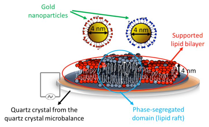
“Formation of supported lipid bilayers…” I will not spend too much time on the first part of the title as my friend and colleague Lisa Jacob at the University of Illinois has previously written a blog post that discusses lipids and supported lipid bilayers. Supported lipid bilayers are basically synthetic cell membranes that can be made in the lab. An actual cell membrane is made up of MANY different biological molecules including phospholipids, glycolipids, cholesterol, proteins, lipopolysaccharides, peptidoglycans… I could go on, but I think you get the point. In order to reduce the complexity of real cell membranes we can make supported lipid bilayers in the lab that are made of just a few biological molecules. This allows us to study the importance of different molecules individually. It is often easier to study a simplified system (like supported lipid bilayers), but it does not behave exactly like the membrane of a living cell – unfortunately, there are usually tradeoffs for simplicity.
“…containing phase-segregated domains…” This is where things become new and exciting! Phase-segregated domains are also sometimes referred to as “lipid rafts,” so from here on out I’ll just call them that (simplicity again!). Lipid rafts are small regions within a cell membrane where the lipids are more tightly packed and the combination of different types of lipids is different than in the majority of the cell membrane. They function like platforms that allow particular cell functions to occur. Remember the movie Castaway when Tom Hanks was floating on a raft in the sea? You can think of lipid rafts similarly, but instead of Tom Hanks there are specific biological molecules that like to be associated with lipid rafts. Another interesting thing about lipid rafts is that they are found in the cell membranes of many different organisms, from plants to animals to bacteria. Many scientists study lipid rafts to understand their importance for living cells.
Finally getting to the first interesting finding in this paper, we were the first people to explain how to make supported lipid bilayers that contain lipid rafts that can be studied with an instrument called a quartz crystal microbalance. Another good friend, colleague, and co-author on this publication, Arielle Mensch from the University of Wisconsin, wrote a blog post explaining this instrument. As scientists, we always want to be VERY confident in our results before publishing them, so we confirmed the presence of these lipid rafts using both the atomic force microscope (see Figure 3 below) and the incredible super-resolution fluorescence microscopy techniques available at the Pacific Northwest National Laboratory (the place I now call my scientific home!). (See our blog posts here and here for more on super-resolution microscopy.)
This was an important finding because, as I mentioned before, many scientists study lipid rafts, and the quartz crystal microbalance instrument provides a new way to study the dynamic biological processes occurring at these special sections of the membrane.

“…and their interaction with gold nanoparticles” At the Center for Sustainable Nanotechnology our primary goal is to investigate the fundamental molecular mechanisms by which nanoparticles interact with biological systems. Through these investigations we hope to redesign nanoparticles that are both useful and environmentally safe. In the case of this publication, we didn’t just use the quartz crystal microbalance instrument because no one had done it before. We used it to figure out how the presence of lipid rafts in supported lipid bilayers impacts the interaction of cell membranes with gold nanoparticles. We found that, under some conditions, the presence of lipid rafts results in more positively-charged gold nanoparticles attaching to the supported lipid bilayers than when there were no lipid rafts. As I said, lipid rafts are found in the cell membranes of many different organisms, so this has potentially broad implications for understanding what happens when gold nanoparticles come into contact with cells.
One of the many beauties of science is that the results of our experiments almost always lead us to ask new questions. For example, why do more nanoparticles associate with supported lipid bilayers that have lipid rafts? Will we observe the same result in supported lipid bilayers that contain more of the complex biological molecules that are found in actual cell membranes? Does the type of nanoparticle matter, or will all positively-charged nanoparticles show the same result as gold? What would this increase in attachment of nanoparticles mean for a living cell? We will be working hard to answer these and many other questions, so stay tuned to our blog for breaking details!
This post is part of our ongoing series of public-friendly summaries describing research articles that have been published by members of the Center for Sustainable Nanotechnology. This collaborative study was done by researchers from the University of Wisconsin-Madison, Pacific Northwest National Laboratory, and University of Illinois at Urbana-Champaign. Eric Melby, a doctoral student at UW-Madison currently based at PNNL, was the paper’s first author. The article was first published online in September 2015 in Environmental Science: Nano.1
EDUCATIONAL RESOURCES
- TedEd animation: Insights into cell membranes via dish detergent – Ethan Perlstein
- Several cell activities by Ellen McHenry, including a fluid mosaic model, human membrane group activity, and 3D membrane paper model
- Melby E., Mensch A., Lohse S., Hu D., Orr G., Murphy C., Hamers R., & Pedersen J. Formation of supported lipid bilayers containing phase-segregated domains and their interaction with gold nanoparticles. Environmental Science: Nano, 2015. doi: 10.1039/C5EN00098J

