One major type of output from university research labs is the publication of scientific results in scientific journals. When we write these papers, our target audience is not the general public; rather, we are writing for experts in our area to tell them what we’ve accomplished so that they can build on our work in their own continuing research. These journal articles are an often-used measure of a university professor or graduate student’s success – people track how many scientific journal articles they’ve published and how many people have cited their papers in ongoing work. From the perspective of the general public, however, scientific journal articles can be difficult to read and digest. That said, they are critical to keep the scientific enterprise moving forward. If everyone kept their experimental results to themselves, much time and money would be wasted as many laboratories unknowingly pursued the same experiments.

In hopes of making a recent Center for Sustainable Nanotechnology paper more relevant to the general public, I’m going to spend this blog post describing recent work and explaining why it’s important. Recently, some of the researchers in our center published a paper titled, “Facile Method to Stain the Bacterial Cell Surface for Super-Resolution Fluorescence Microscopy” in a journal known as Analyst. The title alone can be quite intimidating but, put simply, this paper describes a way that researchers in our center have been able to visualize bacterial cells more clearly than can be done with any standard light microscopes.
The graduate student who led this work, Ian, figured out a way to put a glowing (fluorescent) molecule only on the surface of bacterial cells. This fluorescent molecule bonds to a chemical functional group known as a primary amine, a molecular species that can be found all over bacterial membranes and the membranes of mammalian cells as well. One property of this particular fluorescent molecule is that the glowing blinks on and off quickly. This blinking ends up being critical to achieving very detailed images.
In imaging science, it is critical to be able to tell the difference between two objects that are very close to one another. This is known as “spatial resolution.” This method achieved excellent spatial resolution by taking advantage of the blinking of the fluorescent molecules on the bacterial membrane surface. When molecules are blinking on and off, it is possible to mathematically determine the center of the location of each blinking spot. Ian described this method in more detail in another blog post. For comparison, a traditional light microscope allows a viewer to distinguish objects as small as 250 nanometers in size. In this scientific manuscript, we reported the ability to distinguish objects as small as 30 nanometers, nearly 10 times smaller.
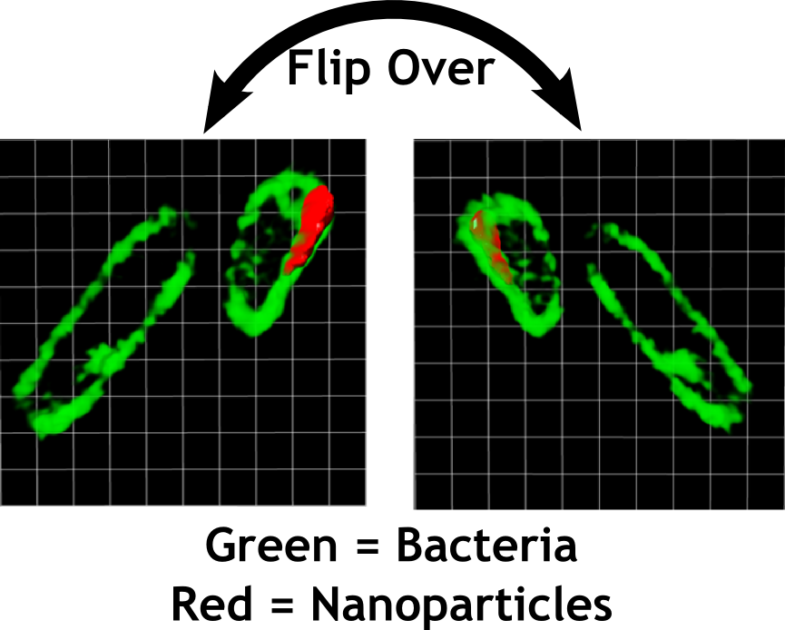
This improved spatial resolution means that it is possible to distinguish bacterial cell walls from the interior of the bacterium, for example. This turns out to be critically important! Of course, in the Center for Sustainable Nanotechnology, we’re interested in how nanoparticles interact with biological organisms so that we understand how to create nanomaterials that are useful and safe. So, now that Ian had shown that we had a very powerful way to visualize bacteria, he had to add the nanoparticles. In this case, he chose a nanoparticle that glows a different color than the molecule on the surface of the bacteria. The type of nanoparticle Ian chose to work with is known as a quantum dot. Because of their strong fluorescent properties, quantum dots are currently being used in ultrabright television and cell phone displays. Ian allowed these quantum dots time to interact with the bacteria and then recorded images on a powerful microscope located at the Pacific Northwest National Laboratory in Washington state. These images revealed that, in some cases, the quantum dots were sticking to the membrane of the bacteria.
Using previous techniques it would have been nearly impossible to understand much more than this. Using this new technique however, we were able to discover that the nanoparticles don’t penetrate the bacterial membrane, but are kept on the outside. Without this specialized, high spatial resolution imaging, it wouldn’t have been possible to tell if the nanoparticles near the bacteria were inside or outside the bacteria, a critical difference when considering how the nanoparticles might impact the bacterial cells. The beautiful (well, beautiful to scientists, at least) images led us to pose new hypotheses about which molecular components of bacteria might be most important in determining any interactions between bacteria and the nanoparticles. More on those hypotheses in a future post.
Pushing the limits of these optical methods led us to better understand how nanoparticles interact with one component of the ecosystem and helped us figure out which scientific questions to ask next. By publishing this work in a scientific journal, we’ve also let other scientists know how they can do high resolution imaging of bacteria for studies with nanoparticles or for other purposes entirely.


[…] research, as well. For example, CSN researchers use fluorescence (though not fluorescein) to detect certain molecules on the surface of bacteria cells. One of our most popular posts explains how fluorescence makes highlighters glow. Because we […]
[…] as small as tens of nanometers. Dr. Christy Haynes talked about super-resolution microscopy in a recent blog post, so I won’t cover that again here. However, one of the enabling technologies for this year’s […]