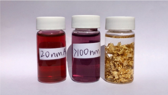
In a previous post, Can gold melt at room temperature? Melting temperature depression!, we talked about how the color of gold changes from shiny yellow to dark red when it is shrunk down to the nanoscale. We know that lots of materials have interesting properties when they get that small, but why do gold nanoparticles have such a range of unexpected colors?
To answer this question, we have to understand the nature of light and what makes the colors we see every day. Wave–particle duality is a concept that describes that light and matter exhibit properties of both waves and particles. This might sound at first like I’m describing something as being like both a ball and a slinky at the same time, but wave-particle duality is very useful for understanding how light behaves.

Photons, which are the “particles” of light, can be used to describe the absorption of light from materials. When light energy is absorbed by a surface, it can be converted to other types of energy such as heat or electricity. The reason why we feel warm bathing the sunlight is because we absorbed light (photon) energy emitted from the sun.
Figure 3 shows an example of the famous photoelectric effect that Albert Einstein used to explain light as particles (photons). Photons are particles and contain energy. Photons in blue light have higher energy than photons in red light, so when the metal plate absorbs blue light photons, it gets enough energy to emit electrons and release the extra negative charge. That negative charge causes the metallic foil inside the bottle to start to fold when blue light hits it. Red light, on the other hand, cannot make the metallic foil folds because of lower photon energy. This simple experiment allowed scientists to demonstrate how light energy works without needing any fancy microscopes or other expensive equipment.

The other half of the particle-wave duality says that light can also be considered as electromagnetic waves. Figure 4 shows an example of an experiment that can only be explained by considering light as waves, called the Young’s interference experiment. This experiment showed that light (electromagnetic waves) passing through two slits (double-slit) add together or cancel each other out, which causes interference fringes to appear, similar to what we would observe for water waves.

In a broad sense, “light” can refer to electromagnetic waves of any wavelength, no matter if they are visible or not. The visible spectrum is the portion of light that is visible to the human eye, typically from about 380 to 740 nanometers. Any colors we see are essentially the result of the selective reflection and absorption of the light source. This is most commonly white light, which contains all the wavelengths of the visible spectrum at almost equal intensity (e.g. daylight or white LED lights). When one wavelength of light is absorbed, the light that reflects most strongly back to our eye tends to be “complementary,” shown on the color wheel in Figure 5. (For more on light waves and how we see color, check out this blog post.)

For example, grass is green because the chlorophyll in the grass absorbs strongly in the red and blue spectrum of white light and reflects the rest of the spectrum, which is mostly green (Figure 6).

Metals, such as the gold that launched our quest for understanding color changes, are generally good thermal and electrical conductors, which means that heat and electricity can travel through them without changing very much. This is because the metal ions are closely packed together, and have many electrons that can carry kinetic energy through these closely packed molecules. These electrons in gold nanoparticles (of roughly 5 – 300 nm in diameter) can respond to the incoming light with something called localized surface plasmon resonances (LSPRs). LSPRs are wavelengths of light where certain electrons in the gold resonate with the incoming light waves, so that the nanoparticle can absorb the incoming light and those electrons actually end up moving based on where the peaks and valleys of the light waves are. (Figure 7)

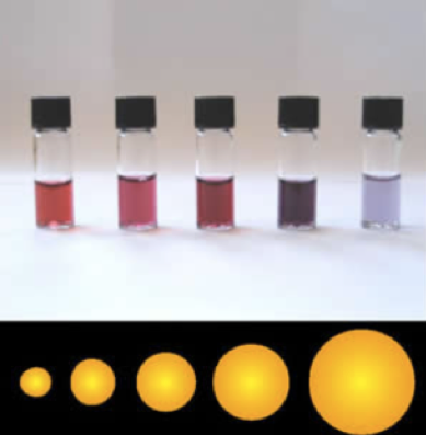
The LSPR of 5-10 nm gold nanospheres ranges from 520 to 580 nm, which means that they absorb green or yellow light. As a result, they show the complementary color of green or yellow, which is red or purple (Figure 8).
While the optical properties of gold nanospheres are determined by a single LSPR, gold nanoparticles shaped like rods get more complicated. They have two LSPRs: one located at around 520 nm corresponding to their short axis, and the other located at higher wavelengths corresponding to their long axis (Figure 9).

The exact wavelength of this higher LSPR depends on the length-to-width ratio of the nanorods. This means that by making rods of different sizes, we can tune the higher LSPR from 600 nm into the near infrared region, so that gold nanorods of different aspect ratios show a “rainbow” of colors (Figure 10). Similarly, other shapes, such as gold nanostars or gold nanourchins,3 also have distinct colors.

Other than giving gold nanoparticles their color, the unique plasmonic properties of metal nanomaterials are also very useful for other applications. Raman spectroscopy is a great analytical technique to determine molecular structures, but it suffers from weak signal intensity. We can use the plasmonic properties of gold nanoparticles to enhance the Raman signal (called surface enhanced Raman spectroscopy), which can even allow single molecule detection. That could be a good topic for another blog post!
EDUCATIONAL RESOURCES
- NASA lesson plan: Discovering Color with a Prism (grades 9-12)
- Veritasium video: The Original Double Slit Experiment
REFERENCES
- Abadeer, N. S., & Murphy, C. J. Recent progress in cancer thermal therapy using gold nanoparticles. The Journal of Physical Chemistry C, 2016, 120(9), 4691-4716. Doi: 10.1021/acs.jpcc.5b11232
- Burrows, N.D., Lin, W., Hinman, J.G., Dennison, J.M., Vartanian, A.M., Abadeer, N.S., Grzincic, E.M., Jacob, L.M., Li, J. and Murphy, C.J. Surface chemistry of gold nanorods. Langmuir, 2016, 32(39), pp.9905-9921. Doi: 10.1021/acs.langmuir.6b02706
- Bakr, O.M., Wunsch, B.H. and Stellacci, F. High-yield synthesis of multi-branched urchin-like gold nanoparticles. Chemistry of Materials, 2006, 18(14), pp.3297-3301. doi: 10.1021/cm060681i
- Burrows, N. D., Harvey, S., Idesis, F. A., & Murphy, C. J. Understanding the seed-mediated growth of gold nanorods through a fractional factorial design of experiments. Langmuir, 2017, 33(8), 1891-1907. Doi: 10.1021/acs.langmuir.6b03606

