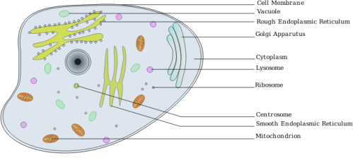
One of the primary goals of the Center for Sustainable Nanotechnology is to understand what happens when nanomaterials are exposed to biological systems like organisms or even cells. How can we do this? You may think back to an introductory biology class and recall a few buzz words about cells, like endoplasmic reticulum, vesicles, and cell membrane. All of these different components serve important functions within the cell, but the cell membrane (the outermost part of the cell) is the key to understanding how the cell interacts with nanoparticles. The cell membrane can even inform us about more complex interactions, such as between nanoparticles and whole organisms.

The main purpose of the cell membrane is to act as a barrier to protect the different components of the cell from the external environment. The cell membrane also allows for transport of cell materials to and from the cell. When nanoparticles are exposed to cells, it is possible for them to interact with the membrane and alter how it functions.
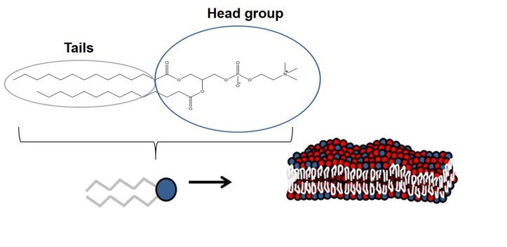
To study this interaction, we need to develop an approximate model of the cell membrane surface. The cell surface is a membrane consisting of a phospholipid bilayer with embedded proteins. Phospholipids (sometimes abbreviated as just lipids) are naturally-occurring chemical compounds that have water-loving (hydrophilic) heads and water-hating (hydrophobic) tails. When the lipids cluster together with their tails away from water and their head groups pointed toward water, they form a bilayer.

By forming bilayers, the tail groups are shielded from water by the external head groups. This creates the barrier that protects the internal parts of the cell from the outer environment. There are also various proteins wedged into the bilayer which serve different functions. For example, certain proteins are responsible for assisting in communication between the cell and the external environment.
With this understanding of the cell membrane, we can model different bilayer structures in the laboratory to study how they would interact with various nanoparticles. The structures can be flat and formed on a surface that serves to anchor the bilayer, called a supported lipid bilayer (SLB). Or it can be spherical like lipid vesicles, called liposomes.

We can design a great variety of SLBs or liposomes by choosing from a range of different lipids. A number of different lipids are possible because each lipid can have a distinct tail and head group. The lipids can also be fluorescent so that we can do cool imaging experiments that highlight the bilayer structures. We can also insert components like membrane proteins into the bilayer structures. The versatility in designing these systems allows us to do well-controlled studies that simulate the cell, which helps us determine specifically what factors affect nanoparticle/cell interactions. Many researchers in the Center study the effects of nanomaterial exposure on these systems.
Experiments on these simplified model membrane systems provide key information for understanding not only single cells, but will also be useful for studying how nanomaterials interact with whole organisms! With this knowledge, we would be able to anticipate how a given nanomaterial will affect an organism, allowing for the design of nanomaterials that are safe and non-toxic.
If you want more information on lipids and cell membranes, here are a few links:
Chem4Kids
Virtual Chembook
Carnegie Mellon Biology Animations

[…] to track individual lipid molecules diffusing within a model cell membrane (specifically, a supported lipid bilayer) with and without nanoparticles present. This is absolutely […]
[…] of the different nanomaterials that we use, but I mainly use it to study the interactions between supported lipid bilayers (a mimic for cell membranes) and various nanoparticles. One benefit of AFM is that we can perform […]
[…] I quickly understood the far ranging possibilities of this research, as all living organism have lipid bilayers; I was sold on the […]
[…] Another important note is that often the methods one uses to study something are as important as the focus of study itself. Our research was done as part of the CSN’s effort to improve the approach for how nanomaterials research is conducted. Doing so required combining the synthetic prowess of the Murphy and Hamers labs and can provide great insight into the CSN’s biological experiments such as those that study water fleas or lipid bilayers. […]
[…] system, we used a laser technique called second harmonic generation to determine that the model, a lipid bilayer, is negatively charged. Given that, we expected that positively charged nanoparticles would […]
[…] synthetic cell membrane that is designed to function like the outside of an actual human cell. (See last week’s blog post for more […]