Here is the underside of a ladybug. Click the image to enlarge. You won’t regret it!

In this post I hope to help you appreciate just how small “nano” is, using the official ladybug of science!
Before we get there, let me explain how I took this image.
Step 1 – Find a dead ladybug. As I am the bug-loving sort, I generally try not to kill them myself. I found this one hanging out in the glass casing underneath a ceiling light during spring cleaning.
Step 2 – Coat the dead ladybug in a thin layer of pure gold. This intensifies how crazy I sound, but is also a critical step in acquiring images like this using a high-powered microscope known as a scanning electron microscope (SEM). This microscope uses electron waves instead of light waves. The bug needs to be coated with gold so the electrons can be conducted away from the area of investigation. Conductive materials or surfaces are critical to obtaining any image using a microscope of this kind.
Step 3 – Put a few drops of nanoparticle solution on the ladybug. I did this so when we zoom in on this image, we can see just how small nanoparticles are in comparison to this already small ladybug.
Here is what the ladybug looked like after steps 1 through 3.

Step 4 – Put the ladybug in the scanning electron microscope. See a future post for more detail on this step.
Step 5 – Acquire a series of images and spend a few hours at home with your dog of science, painstakingly stitching the images together into the big composite image you see at the top of this post.
Now here comes the really fun part. Since this ladybug is coated with nanoparticles, we can zoom in to see just how much smaller the nanoparticles are than this already-tiny bug.
Here goes! We’ll start with it’s little foot-looking-part. The nanoparticles will be little spheres—either light- or dark-colored depending on how zoomed in we are.


Look at all those nanoparticles!! Well, technically they’re not “nano” particles, as they are around 400-500 nanometers in diameter, and “real” nanoparticles need to be less than 100 nanometers in diameter. Here is a mega-zoom-in of a few nanoparticles. These nanoparticles are made from silica, just like sand. Nano sand grains!
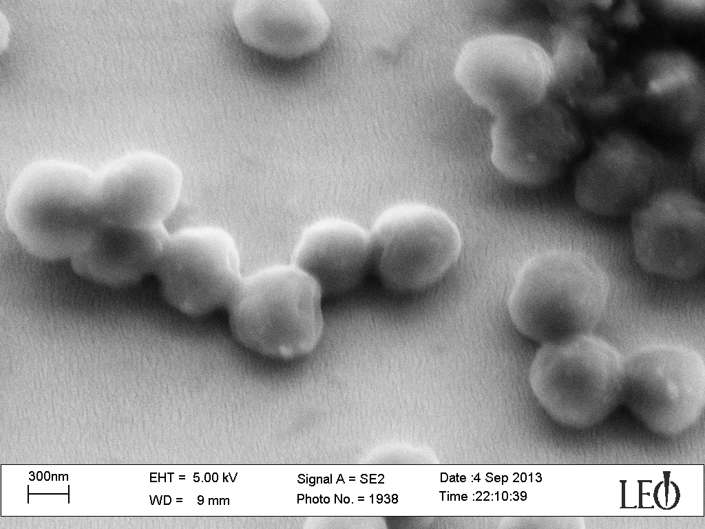
The particles in this image are almost 100 times larger than the nanoparticles I use for my research!
So, as promised in the title, here is the animated version of the above images:

As you can see, nanoparticles are SMALL!! Since you have almost no hope of diverting your eyes away from the animation above, I’m just going to finish here and tell you to stay tuned for future posts about how these amazing microscopes work and how we use them for more standard research purposes.
Related Activities
Exploring Size: Powers of Ten
Exploring Size: Memory Game
Life Size: Line ’em Up

